Structure of Skeletal Muscles
Skeletal muscles are made up of a collection of muscle cells, or muscle fibers, which are long, cylindrical cells with hundreds of nuclei on their surface, arranged in parallel. The main intracellular structures of the muscle cells are myofibrils, the contractile units of the cells which are composed of organized bundles of contractile and elastic proteins[40].
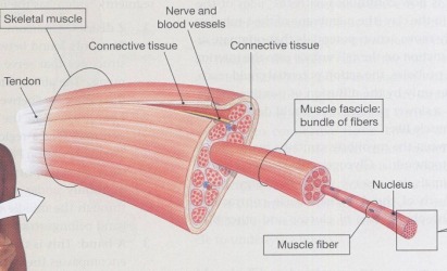
Myosin and actin are the contractile proteins, also known as the thick (myosin) and thin (actin) filaments. Thick filaments are made of around 250 myosin molecules, which are in turn composed of intertwined protein chains that form a long tail and a pair of heads. Thin filaments by contrast are composed of two chains of actin polymers twisted together into a double strand. The parallel thick and thin filaments are connected via crossbridges that form when myosin heads bind loosely to actin subunits of the thin filaments. During the ATP-dependent contractile cycle of a muscle fiber, myosin heads ratchet along the actin filaments, causing the entire fiber to shorten[40].

The regulatory proteins in a myofibril are troponin and tropomyosin. The latter is an elongated polymer that wraps around the actin filament and partially blocks the myosin binding sites, preventing myosin from completing
its contractile cycle. Tropomyosin is slid across the surface of the thin filament to allow complete binding of myosin by troponin. This shift occurs when calcium ions bind to troponin[40].

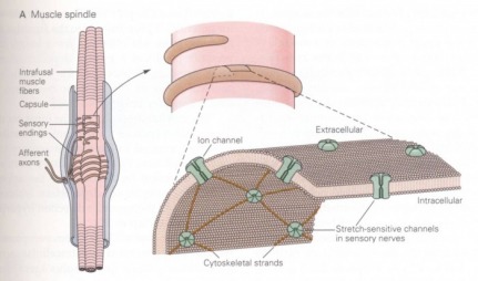
Muscle spindles are small elongated stretch receptors scattered among the contractile fibers. They are responsible for sending information about muscle length to the central nervous system. Each spindle consists of a capsule of connective tissue enclosing intrafusal fibers. These are muscle fibers which have been modified so that only the ends are contractile. These ends are innervated by a gamma motor neuron. The noncontractile center region is wrapped by sensory nerve endings which project to the spinal cord and synapse directly on the alpha motor neurons innervating the muscle in which the spindles lie[40].
Picture References
Pictures from course notes.
