Nerve Transferal Surgery
The goal of the surgery is to perform a set of nerve transfers from divided nerves of the amputated limb to otherwise functionless muscle in order to biologically amplify the weak nerve action potentials into much stronger electromyographic signals. An example of this is transferring the divided nerves that enervated an arm into the chest muscle. With the biological amplification of the signal, muscle activity, under voluntary cortical control, is able to guide the activity of a myoelectric prosthesis. The researchers who developed the process define a myoneurosome as a muscle segment under voluntary cortical control with an identifiable vascular and nerve supply that is isolatable by electromyography from other surrounding muscles. The procedure is designed to create new myoneurosomes via nerve transfers. In order to allow for better recording of electromyographic signals on the skin surface, the skin over each myoneurosome is aggressively defatted[23].
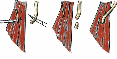
Diagram of nerve transfer. (Left) Intact myoneurosome is on the left. A large nerve with end neuroma is
on the right. (Center) A small motor nerve to the myoneurosome is cut 1 cmfrom its entry point into the muscle.
The neuroma is excised back to healthy fascicles for the large peripheral nerve. (Right) Anastomosis of the large
peripheral nerve to the muscle segment at the entry site of the motor nerve into the muscle. The large peripheral
nerve covers the smaller motor nerve as it enters the muscle[23].
After the nerve brances are identified and tagged, they are debrided back to healthy unscarred fascicles and further proximal dissection is carried out to gain length. The muscle segments are separated by at least two centimeters to keep the different muscle segments’ signals from competing on the electromyogram. Since the muscles are only used as bioamplifiers and not for any type of movement, their repositioning is irrelevant to their function. After the muscle separation, the nerve transfers are performed by sewing the nerve branches to different segments of muscle under loupe magnification. The nerve transfers are performed at the entry points the previous motor neurons had used[23].
